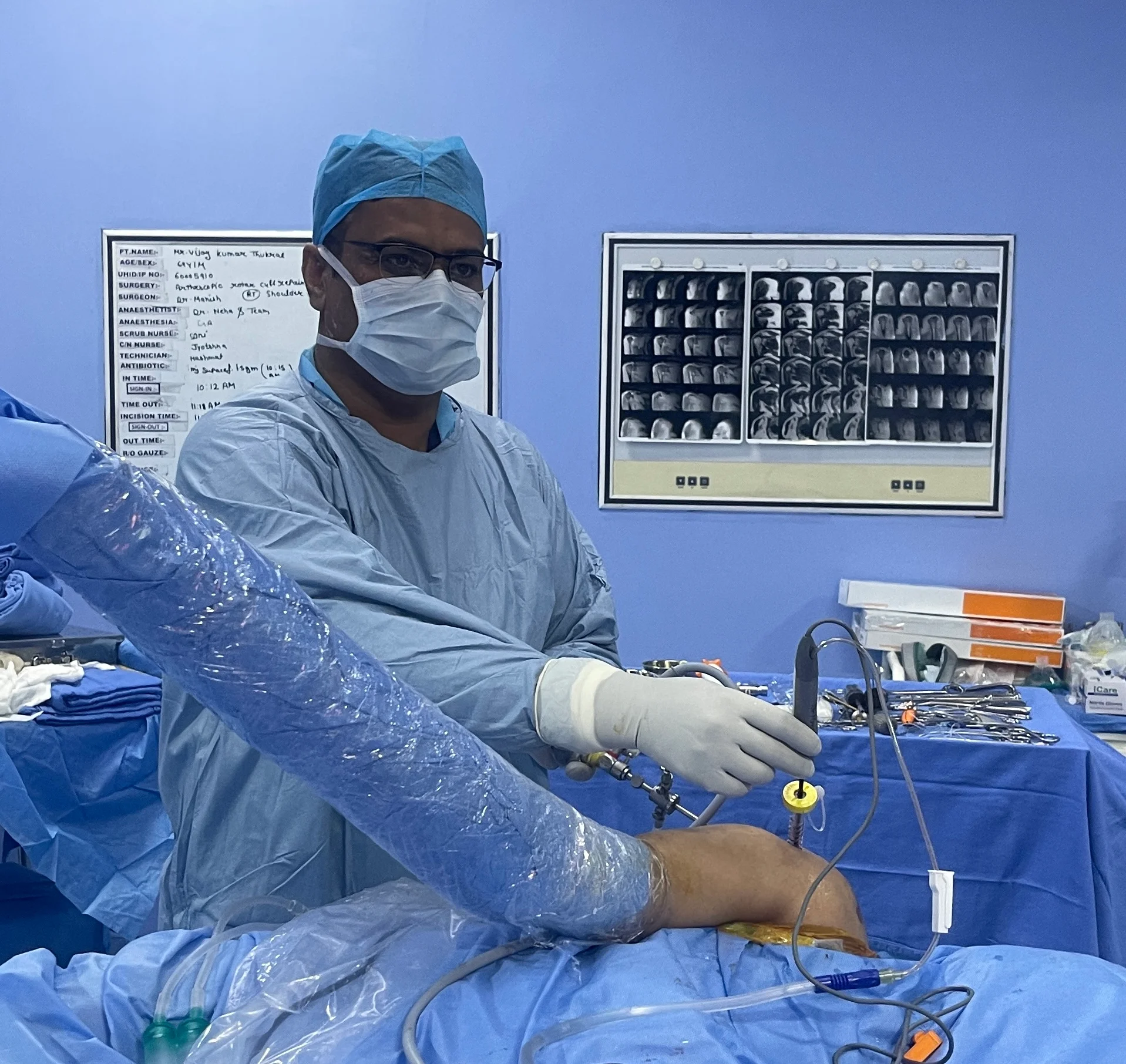
Shoulder Instability Specialist

Recurrent Dislocation of Shoulder

The shoulder joint is the most mobile joint in the human body, allowing a wide range of motion. However, this extensive mobility comes at the cost of stability, making it highly susceptible to dislocation. Recurrent shoulder dislocation is a condition characterized by repeated instances of the shoulder popping out of its socket, which can significantly affect a person’s quality of life and physical activity.

Advanced

Bankart Repair

Minimal

Arthroscopic Care

Recurrent Dislocation of Shoulder Clinical Consultation
Advanced Stabilization
Joint Preservation
Evaluating Shoulder Mobility and Joint Motion
Overview

Recurrent Dislocation of Shoulder Introduction

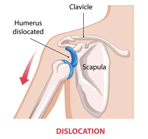
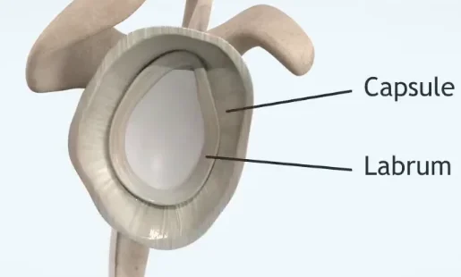
A shoulder dislocation occurs when the upper arm bone (humerus) pops out of the cup-shaped socket (glenoid) that is part of the shoulder blade. The initial dislocation is often the result of a significant trauma, such as a sports injury, fall, or accident. When the forces involved stretch or tear the surrounding ligaments and the labrum (the cartilage ring that deepens the socket), the shoulder loses its primary structural support.

Following a primary dislocation, the damaged structures may fail to heal properly or remain stretched, leading to persistent joint instability. This vulnerability causes subsequent dislocations to happen with progressively less force, sometimes during routine daily movements like sleeping, putting on a coat, or reaching overhead.
Anatomical Evaluation of Shoulder Instability
Pathology

What is Recurrent Shoulder Dislocation

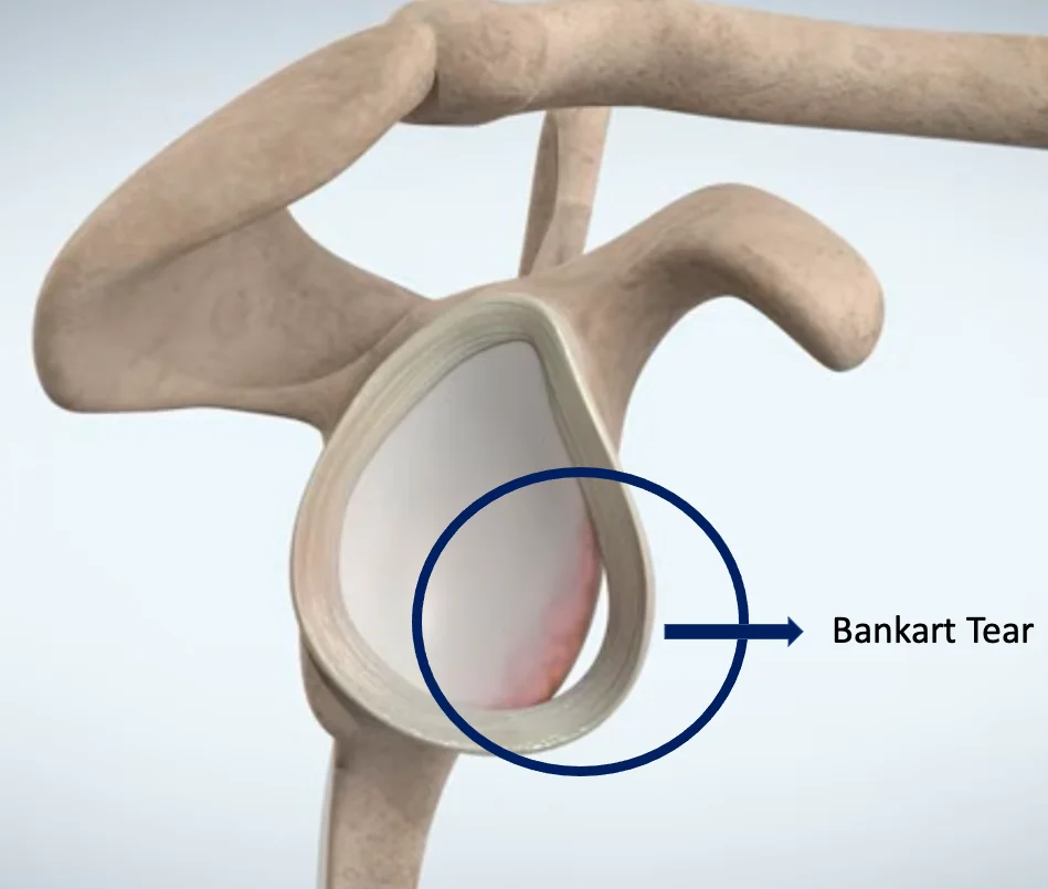
Recurrent shoulder dislocation is defined as chronic shoulder instability resulting from repeated detachments or subluxations of the glenohumeral joint. When a dislocation happens repeatedly, it is usually because the soft tissues that hold the ball in the socket—specifically the labrum and joint capsule—have been torn or permanently stretched out. This tissue damage is commonly known as a Bankart lesion.

In addition to soft tissue injuries, repeated dislocations can lead to bone loss on either the glenoid socket or the humeral head (known as a Hill-Sachs lesion). The loss of these structural bony barriers severely compromises the mechanical stability of the shoulder, necessitating specialized orthopedic intervention.

Etiology

Causes

Understanding the primary mechanisms driving joint laxity is essential to defining the correct path toward permanent stabilization.

01

Severe Initial Trauma

A high-energy primary dislocation—such as from a sports collision, vehicular accident, or a severe fall—can heavily tear the labrum, capsule, and stabilizing ligaments beyond their capacity to heal naturally.

02

Repetitive Overhead Strain

Athletes engaged in repetitive overhead sports like swimming, tennis, throwing, or weightlifting can gradually stretch out the anterior capsule, causing microscopic wear and generalized laxity.

03

Genetic Hyperlaxity

Some individuals possess naturally loose ligaments throughout their bodies due to connective tissue characteristics. In these patients, the shoulder may easily dislocate in multiple directions with minimal or no trauma.

Identifying Chronic Symptoms and Instability Signs
Symptomatology

Symptoms

The presentation of chronic shoulder instability involves distinct mechanical and subjective symptoms that severely limit function:

  • A persistent feeling that the shoulder is loose, slipping, or "giving way" during specific movements
  • Repeated episodes where the upper arm bone visibly and painfully exits the socket entirely
  • Intense, acute pain and muscle spasms immediately following a subluxation or total dislocation
  • Localized swelling, bruising, and a completely altered visual contour of the shoulder profile
  • Numbness, tingling, or localized weakness in the arm if the displaced bone presses against regional nerves
Diagnostic Framework

Diagnosis

A multi-step evaluation protocol is required to accurately map out both soft tissue damage and potential bone deficits.

Step 01

Clinical Instability Tests

Your orthopedic specialist will perform specific physical maneuvers, such as the Apprehension Test and Load and Shift Test, to check for laxity, direction of instability, and muscle guarding.

Step 02

Standard X-rays

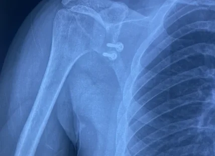
X-rays are critical to confirm a current dislocation state, evaluate general alignment, and initially screen for macro bone defects on the humerus head or glenoid rim.

Step 03

MRI Arthrogram or CT Scan

An MRI (often with contrast dye) provides detailed views of labral tears (Bankart lesions). A CT scan is utilized if significant bone loss is suspected on the socket, helping plan precise bone-grafting procedures.

Expert Insights

Understanding Shoulder Dislocation & Stabilizing Procedures

Watch this detailed clinical presentation explaining how repeated shoulder dislocations affect the labrum and bone structures, along with an overview of modern arthroscopic techniques designed to successfully secure the joint.

  • Mechanism of Bankart and Hill-Sachs injuries
  • Biomechanical explanation of capsule tightening
  • Phases of healing required for complete recovery
Therapeutic Protocol

Treatment Options

Treatment choices are carefully evaluated according to the patient's age, physical demands, lifestyle goals, and structural damage severity.

Nonsurgical Treatment

Conservative therapy can be considered for low-demand patients, or cases strictly driven by generalized multi-directional ligament laxity.

  • Activity Modification: Adjusting daily workflows and avoiding high-risk overhead or contact sports to minimize subluxation triggers.
  • Targeted Physical Therapy: Executing rigorous exercises focused entirely on strengthening the rotator cuff and periscapular muscles to dynamically stabilize the joint.
  • NSAIDs: Utilizing anti-inflammatory medications to decrease post-episode synovitis and joint irritation.

Surgical Treatment

Surgical stabilization is highly recommended for young active individuals, athletes, and anyone experiencing regular dislocations despite undergoing therapy.

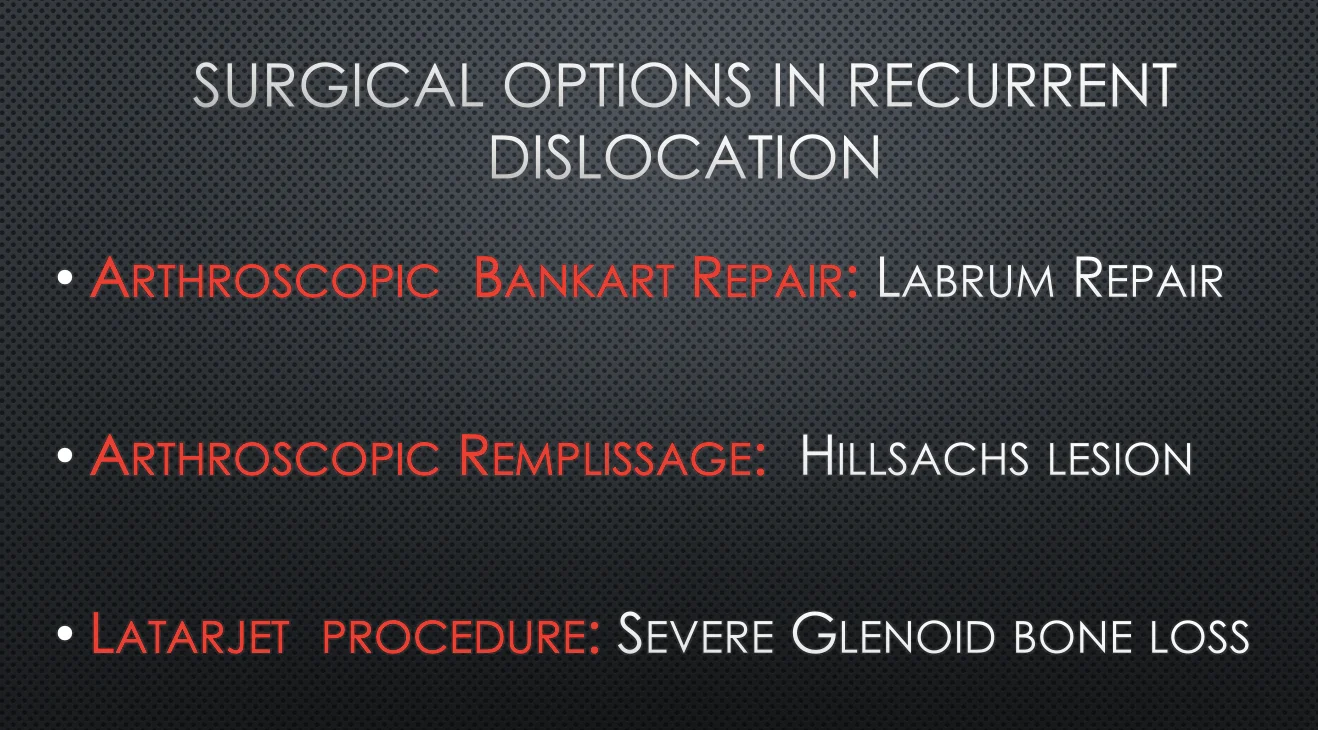
  • Arthroscopic Bankart Repair: Using minimally invasive techniques to reattach the torn labrum back to the glenoid bone using specialized anchors.
  • Open Capsular Shift: An open procedure designed to tighten a significantly stretched joint capsule when tissue laxity is severe.
  • Latarjet Procedure: A bone transfer surgery indicated when substantial bone loss exists on the glenoid socket rim.
Arthroscopic View of Shoulder Labrum Repair
Advanced Surgery

Arthroscopic Shoulder Stabilization / Surgery

Arthroscopic stabilization is the gold standard approach for correcting recurrent anterior shoulder instability. The procedure is performed through a few small, cosmetic incisions around the shoulder joint. A high-definition camera (arthroscope) allows the surgeon to thoroughly inspect the entire joint cavity, identifying the exact pattern of soft tissue tearing and any associated osseous injuries.

Using specialized instruments, the scarred and displaced labrum is carefully mobilized away from its abnormal position. The bone bed on the glenoid rim is prepared to promote bleeding, which is vital for long-term biological bone-to-tendon healing. Small biocompatible suture anchors are then structurally placed into the glenoid edge. The high-strength sutures are woven through the detached labrum and capsule, pulling the tissues back to their anatomical position and effectively restoring the protective bumper ring that stops the ball from sliding out.

Post-Operative Shoulder Physical Rehabilitation Exercises
Rehabilitation Phase

Recovery & Rehabilitation

Post-operative care requires deep adherence to a staged protection and strengthening timeline to allow the repaired labrum to integrate securely with the bone tissue:

  • Immobilization (Weeks 1–4): The operated arm is securely held in a specialized shoulder sling to prevent stress on the newly repaired anterior structures.
  • Passive Range of Motion (Weeks 4–8): Physical therapy begins with gentle, controlled passive movements to prevent joint stiffness while carefully avoiding excessive external rotation.
  • Active Strengthening (Weeks 8–12+): Progression toward full active motion and progressive resistance training targeting the rotator cuff and scapular stabilizers.

A structured return to low-impact sports typically occurs around 4 to 5 months, while full clearance for high-demand contact sports or competitive throwing requires 6 months of comprehensive mechanical recovery.

Patient Guide

Frequently Asked Questions

Review clear, clinically accurate answers to the most common questions regarding chronic shoulder instability management.

The primary shoulder dislocation often tears the labrum cartilage and stretches the joint capsule (Bankart lesion). Because these tissues have limited intrinsic blood supply, they frequently fail to heal back to their normal anatomical positions, leaving the joint inherently loose and susceptible to further dislocations with minimal force.
Physical therapy can strengthen the surrounding muscles to provide dynamic support, which may suffice for low-demand individuals or multi-directional laxity cases. However, therapy cannot structurally repair a physically torn labrum or correct significant bone loss, making surgery the definitive solution for true mechanical instability.
An arthroscopic repair uses small incisions and suture anchors to reattach your native soft tissue (labrum). A Latarjet procedure is an open surgery where a piece of bone from the coracoid process is transferred to the front of the glenoid socket to compensate for severe bone loss that soft tissue repair alone cannot fix.
Arthroscopic Bankart stabilization has a highly favorable track record, boasting success rates between 85% to 95% in restoring joint stability and preventing further dislocations, provided there is no significant pre-existing bone loss and the patient fully complies with post-operative physical rehabilitation protocols.

Restore Your Shoulder Stability Permanently

Break the cycle of recurrent shoulder dislocations and regain absolute confidence in your movements. Schedule a comprehensive diagnostic consultation with our sports medicine experts today.