Advanced Hip Preservation & Joint Care

Avascular Necrosis of Hip (AVN Hip)

Avascular Necrosis of the Hip is a serious joint disease characterized by a temporary or permanent loss of blood supply to the femoral head, leading to structural damage and progressive bone breakdown.

Preserve

Joint Architecture

Targeted

Core Decompression

Avascular Necrosis of Hip Main Interface Design
Joint Preservation
Advanced Arthroplasty
Clinical Identification of AVN Hip Progress
Introduction

AVN Hip Introduction

Avascular Necrosis (AVN) of the hip, also known as osteonecrosis, is a painful condition that occurs when the blood supply to the head of the femur (thighbone) is disrupted. Because bone cells require a steady supply of blood to remain healthy and regenerate, a lack of blood flow causes the bone tissue in the femoral head to die.

Over time, this process weakens the bone structure, eventually causing it to collapse. Once the smooth, rounded head of the femur loses its shape, the hip joint breaks down rapidly, leading to severe arthritis, intense pain, and significant limitations in physical mobility.

Radiological Architecture of Avascular Necrosis
Mechanism

What is Avascular Necrosis of Hip

Avascular Necrosis of the hip joint is a progressive disease that primarily targets the ball portion of the ball-and-socket hip joint. When blood flow is restricted, tiny microscopic breaks develop within the interior structure of the bone. As the disease advances without intervention, these internal micro-fractures compromise the structural integrity of the femoral head.

The bone becomes fragile and can no longer support the weight of the body. When the bone collapses, the overlying articular cartilage loses its foundational support, warping the joint surface and triggering highly debilitating degenerative joint patterns.

Anatomy of the Hip Joint and Femoral Head Structure
Joint Biomechanics

Anatomy of the Hip Joint

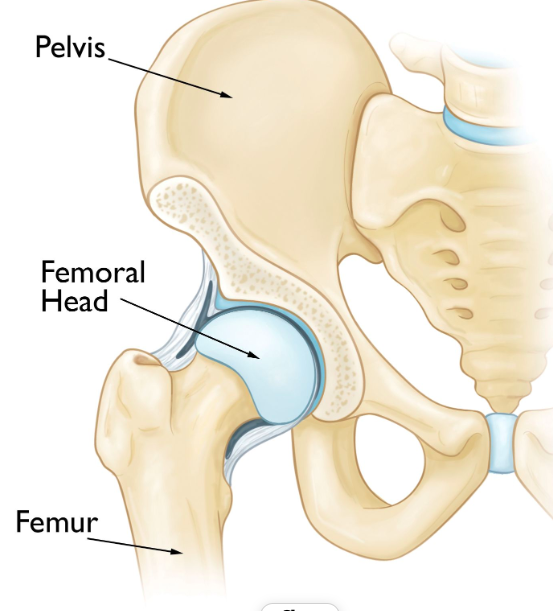
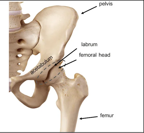
To understand AVN, it helps to understand the natural configuration of the hip joint. The hip is one of the largest weight-bearing ball-and-socket joints in the body. The socket is formed by the acetabulum, which is part of the large pelvis bone, while the ball is the femoral head, located at the upper end of the thighbone.

A delicate network of small blood vessels delivers the essential blood supply required to nourish the femoral head. Because this blood supply is uniquely specialized, any trauma or structural blockage that narrows or cuts off these vessels directly starves the bone cells, setting the path for localized bone death.

Etiology

Causes of AVN

AVN can result from a single acute injury or develop gradually from underlying medical conditions or prolonged systemic factors:

01

Trauma & Dislocation

Hip fractures, severe joint dislocations, or surrounding physical injuries can damage, tear, or compress the delicate blood vessels supplying the femoral head.

02

Corticosteroid Use

High-dose or prolonged use of systemic steroid medications can alter lipid metabolism, increasing fat deposits in the bone marrow that compress local blood flow.

03

Alcohol Consumption

Excessive long-term alcohol intake can lead to fatty substances collecting within the blood vessels, blocking the microcirculation feeding the joint tissue.

Surgical Mapping of Femoral Progression Stages
Disease Progression

Stages of AVN

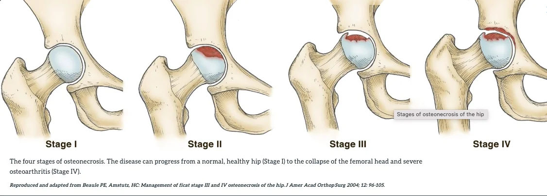
Avascular Necrosis of the hip typically progresses through four primary clinical stages:

  • Stage 1: Early stage where bone changes are silent and invisible on regular X-rays, detectable only via sensitive MRI scans.
  • Stage 2: Clear structural alterations are visible on standard X-rays, but the rounded contour of the femoral head remains fully intact.
  • Stage 3: Structural failure occurs, leading to the collapse of the subchondral bone and creating a flattened or irregular joint line.
  • Stage 4: Severe, widespread degradation of the joint surfaces, culminating in secondary osteoarthritis as the socket cartilage erodes.
Symptomatic Limitation of Pelvic Mobility
Clinical Indicators

Symptoms

In its initial phases, AVN of the hip may cause no discernible symptoms. However, as bone destruction advances, specific physical issues emerge:

  • Mild, intermittent pain inside the groin, thigh, or buttock region
  • Severe, throbbing joint pain that intensifies when putting weight on the affected leg
  • Persistent discomfort that continues even while resting or lying down in bed
  • A progressive reduction in the hip's overall range of motion, making bending or rotating difficult
  • The development of a noticeable limp when walking due to worsening structural mechanical instability
Diagnostic Assessment

Diagnosis & Evaluation

Catching AVN early is vital to preserving the natural joint structure before the femoral head undergoes irreversible collapse.

Initial Screening

X-Ray Examination

Standard plain radiographs are helpful for identifying mid-to-late stage bone changes, showing increased bone density or flattened surfaces characteristic of structural failure.

Advanced Diagnostics

Magnetic Resonance Imaging (MRI)

MRI remains the gold standard for early AVN detection. It can clearly capture initial blood flow deficits and cell death when standard X-rays still appear normal.

Orthopedic Education

Biomechanical Presentation of AVN Hip Joint Pathology

Watch this comprehensive, professionally prepared video presentation exploring the precise clinical mechanisms of Avascular Necrosis of the hip. Learn how structural blood supply restrictions impact the femoral head and see how early surgical preservation strategies contrast with modern arthroplasty procedures.

  • Vascular compromise inside the femoral neck
  • Subchondral bone failure and mapping
  • Prosthetic engineering for anatomical restoration
Therapeutic Planning

Treatment Options

The choice of treatment depends heavily on the specific disease stage, the size of the necrotic bone region, and the patient's age and activity demands. Treatment pathways are broadly categorized into early joint preservation or advanced joint replacement.

Hip Preservation Surgical Modality Application
Preservation Surgery

Hip Preservation Procedures

When AVN is identified early (Stages 1 and 2), before structural shape loss occurs, surgical preservation procedures are utilized to stimulate new bone growth and restore blood flow:

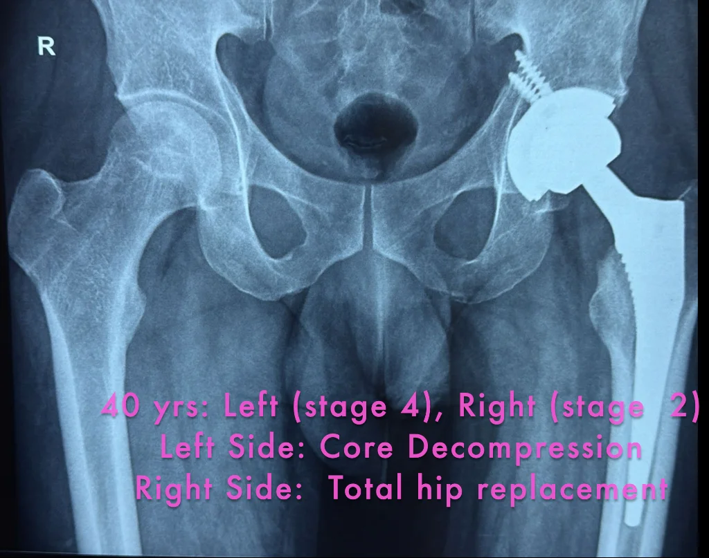
Core Decompression: A widely used procedure where the surgeon drills small channels into the dead area of bone. This relieves internal pressure, creates space, and encourages the growth of new healthy blood vessels.

Bone Grafting: Healthy bone tissue harvested from another part of the body, or a specialized synthetic matrix, is transplanted into the decompressed area to provide solid structural support while the joint remodels.

Total Hip Arthroplasty Configuration for Late-Stage AVN
Arthroplasty Delivery

Total Hip Replacement for AVN

If Avascular Necrosis has progressed to advanced structural failure or joint collapse (Stages 3 and 4), hip preservation is no longer viable. In these scenarios, Total Hip Replacement (total hip arthroplasty) becomes the most successful option.

The surgeon removes the damaged, necrotic femoral ball and replaces it with a smooth prosthetic implant composed of biocompatible metal or ceramic components. The worn-out socket is lined with a durable shell to re-establish fluid, friction-free joint movement, delivering predictable pain relief and returning patients to an active lifestyle.

Post-operative physical therapy rehabilitation process
Rehabilitation Pathway

Recovery & Rehabilitation

Post-operative care is highly tailored to the specific type of surgery performed. Following a core decompression or joint preservation procedure, patients must follow restricted weight-bearing protocols using crutches or walkers for several weeks to protect the healing bone structure.

For patients undergoing a total hip replacement, early mobilization is initiated on day one to prevent vascular clots and maintain flexibility. Structured physical therapy is central to both treatment paths, focusing on rebuilding muscle strength around the pelvis, correcting walking alignment, and maximizing joint range of motion.

Patient Guidance

FAQs

Review key responses to primary medical questions concerning Avascular Necrosis development and modern treatment protocols.

In the vast majority of cases, AVN is a progressive condition that does not reverse spontaneously. While very small, non-weight-bearing necrotic lesions can occasionally stabilize with conservative medical management, progressive AVN typically requires timely surgical intervention to prevent structural bone collapse.
Recovery from core decompression requires a period of restricted weight-bearing, usually lasting 6 to 12 weeks, to safeguard the structurally vulnerable femoral head. Complete structural remodeling and a return to regular physical impact activities can take up to 6 months.
While a history of systemic corticosteroid use is a primary cause of AVN development, it does not compromise the exceptional long-term success rates of modern total hip arthroplasty. The prosthetic joint replaces the affected bone entirely, resolving the localized issue.
Magnetic Resonance Imaging (MRI) is the single most accurate and sensitive diagnostic modality for identifying early-stage AVN. It can clearly visualize bone marrow changes and localized ischemia well before any abnormalities can be detected on standard digital X-rays.

Protect and Restore Your Hip Joint Functionality Today

Connect with our expert orthopedic team to design a precise, personalized medical strategy for treating your AVN symptoms and reclaiming comfortable, restriction-free motion.